EndoNeuroSpine
About the procedure
What Are 30° Endoscopic Procedures?
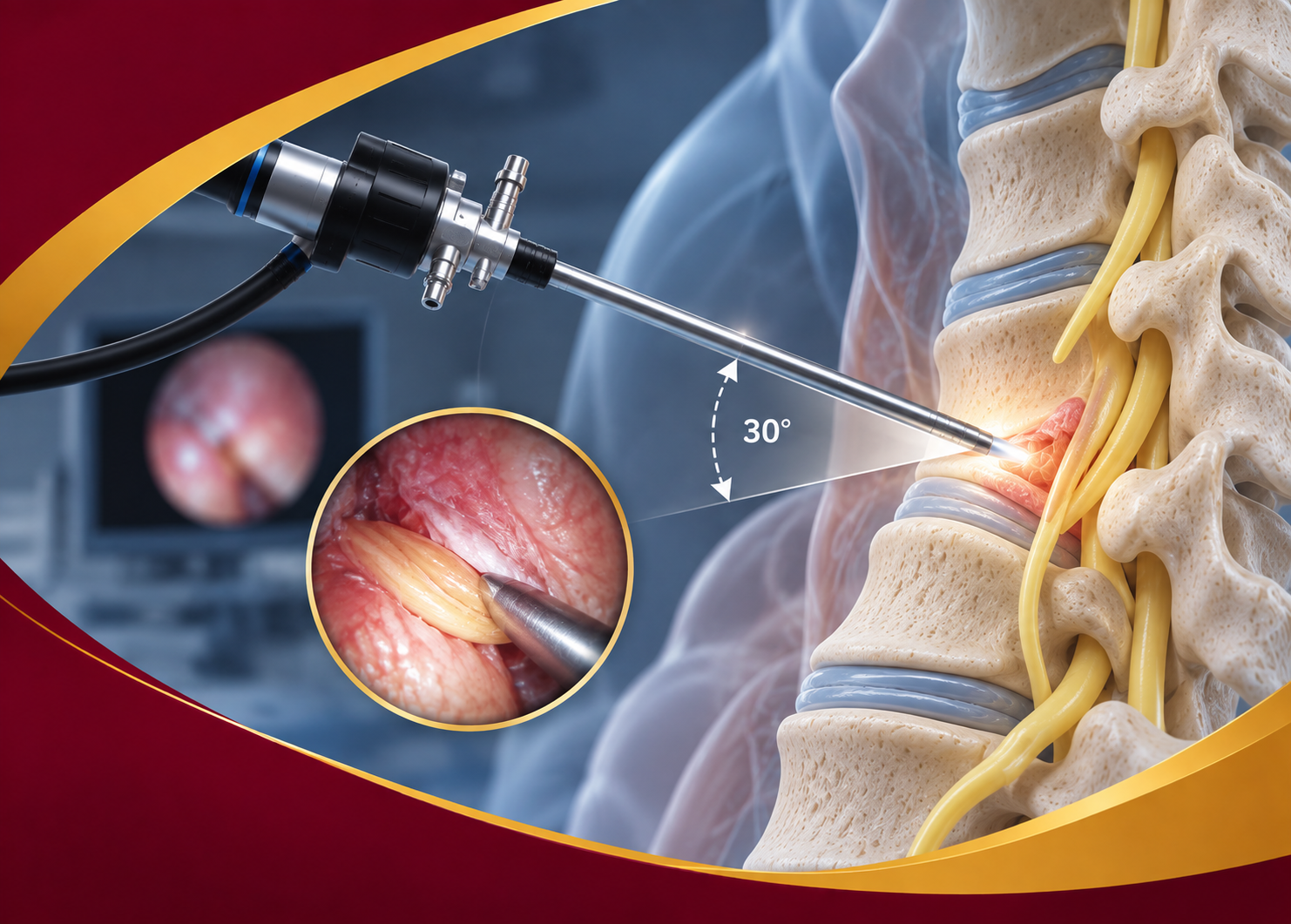
30° Endoscopic Procedures use an angled endoscope so the surgical team can look around anatomy that a straight-on (0°) view may miss, while still working through a small corridor.
The angled lens is designed to widen the field of view in selected brain and spine cases. It does not replace imaging, clinical judgment, or a full discussion of risks. Suitability depends on diagnosis and a specialist’s evaluation.
Conditions we treat
Conditions Where 30° Visualization May Be Considered
30° endoscopic visualization may be considered for selected brain and spine conditions following appropriate clinical evaluation.
Disc Herniation
Spinal Stenosis
Nerve Compression
Confined Anatomy
Other Selected Conditions

Key benefits
The Minimally Invasive Advantage
Potential benefits may include the following, compared with traditional open approaches in appropriately selected cases:
Smaller Incisions
May support a smaller surgical footprint than some traditional open approaches.
Less Pain
Can help reduce tissue disruption for some patients, though discomfort still varies.
Faster Recovery
Some people return to light activity sooner; only your clinician can estimate a timeline.
Better Outcomes
The goal is a plan that may support function and comfort, based on your individual case.
Your treatment journey
How 30° Endoscopic Procedures Work
We follow a personalized approach based on your condition, symptoms, and clinical needs.
1
Consultation
Review symptoms, history, and questions with a specialist.
2
Imaging & Diagnosis
Use imaging and clinical findings to understand whether an angled view may help in your case.
3
Treatment Plan
Discuss whether an endoscopic corridor may be suitable for your case.
4
Procedure
If surgery is advised, treatment uses a small corridor and visualization.
5
Recovery & Support
Follow a written recovery plan and ask before increasing activity.
Our technology
Advanced Visualization for Safer, More Precise Treatment
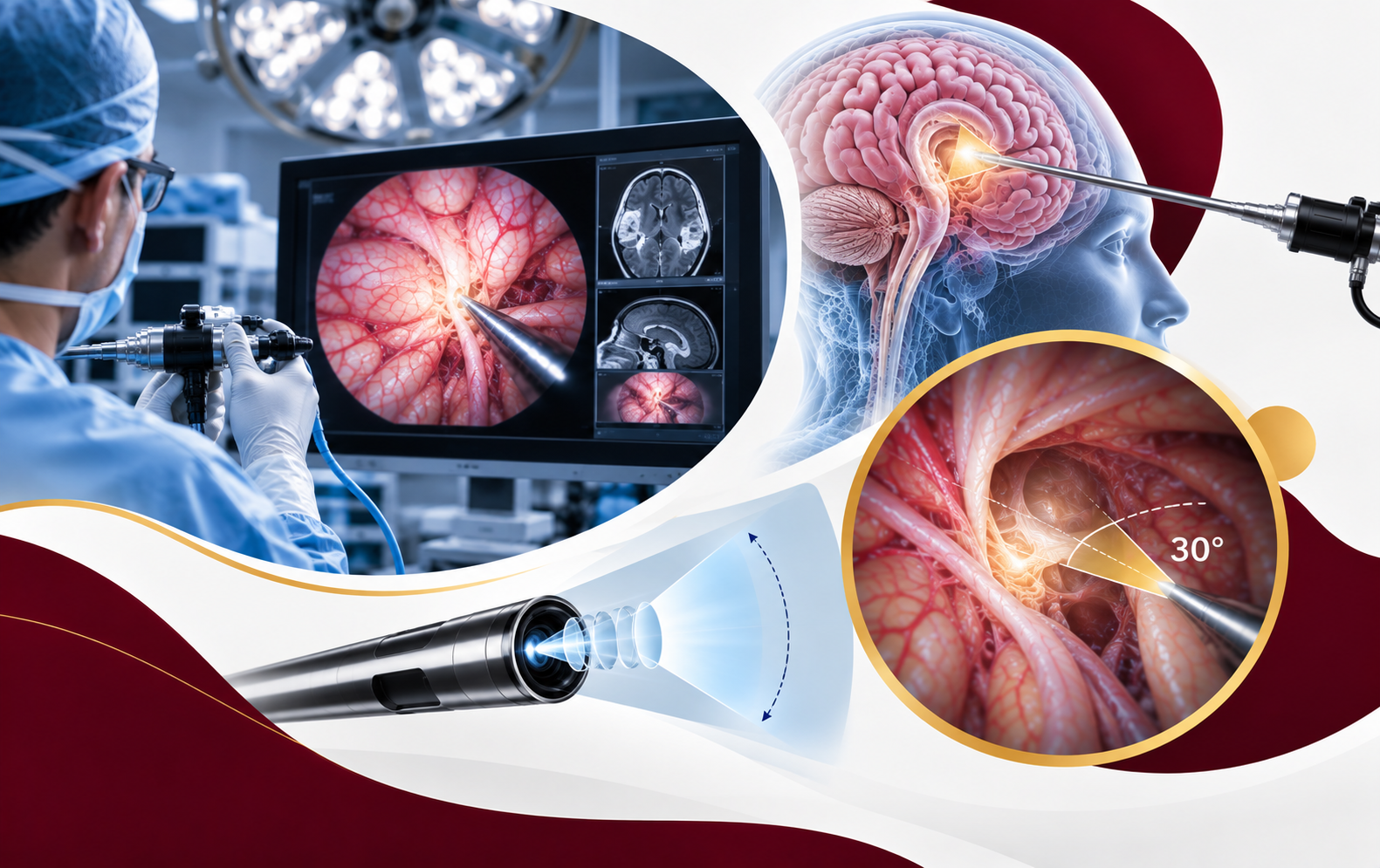
A 30° endoscope is designed to show around structures that sit outside a narrow line of sight. Combined with high-definition imaging, that extra angle can support planning in confined brain or spine corridors.
The lens is a visualization tool, not a guarantee of a smaller incision or a faster recovery. Suitability still depends on imaging, diagnosis, and a specialist’s evaluation. All procedures have risks.
-
High-Definition View
Magnified HD images can help the team see fine detail inside a confined working space.
-
30° Angled Lens
Where used, the angled lens can show around anatomy that a 0° (straight-on) view may miss.
-
Deeper Corridors
Illumination and visualization through a small corridor may support work in selected deep or confined areas.
-
Careful Navigation
Clearer sight lines may support careful handling of nearby tissue. Outcomes still vary from person to person.
 30°
30°
Recovery & results
What to Expect After Treatment
Recovery varies by procedure, condition, and individual patient factors.
Short Hospital Stay
Potentially same-day or next-day discharge depending on the procedure and clinical circumstances.
Gradual Recovery
Return to activities according to individual recovery guidance.
Activity Guidance
Personalized advice for a safe return to work and daily activities.
Long-Term Relief
Follow-up care focused on recovery and functional improvement. Relief is not guaranteed.
Frequently asked questions
Your Questions, Answered.
Here are some common questions about 30° endoscopic procedures. If you have additional questions, please contact our team.
Candidacy depends on symptoms, imaging, anatomy, and overall health. A specialist consultation is needed to determine whether an angled endoscopic view may be appropriate.
No. The angled view can add visual information in selected cases. All surgery has risks. Your care team should explain expected benefits, possible complications, and alternatives. No procedure is risk-free.
Duration depends on the condition, the levels treated, and intraoperative findings. Your specialist can discuss a typical range for your planned procedure. This page does not list a fixed time.
Return to work, exercise, and daily routines is individual. Follow the written plan provided after treatment and ask the clinic before increasing activity.
No. Anatomy, diagnosis, prior surgery, and overall health can mean another approach is safer or more effective. Your specialist will explain alternatives when an endoscopic option is not advised.
Take the next step toward precise visualization
Ask Whether 30° Visualization Applies.
Request a consultation to review imaging and discuss whether a 30° endoscopic approach may be appropriate.